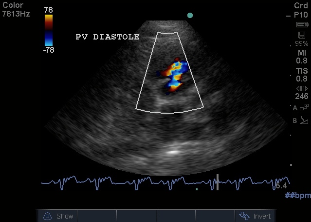
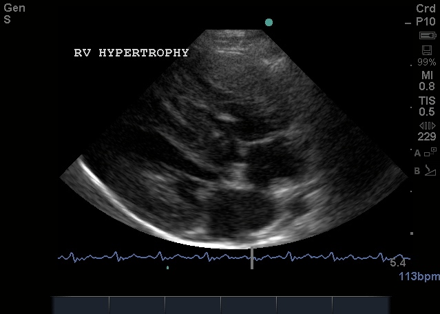
Valvular Pulmonic Stenosis in a 6mo Terrier cross Valvular Pulmonic Stenosis in a 6mo Terrier cross
| CW Doppler trace of Pulmonic Outflow also showing Pulmonic Regurgitation, Same Dog | RV Hypertrophy, Same Dog |
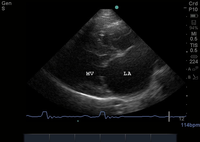
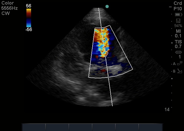
Diastolic frame of Mitral Stenosis in an English Bull Terrier (failure to open) | Colour flow of Mitral Stenosis (Left Apical View) | Continuous Wave Doppler trace of Mitral Stenotic jet, same dog |
Left Parasternal view of Aortic Valve Vegetations | Colour Doppler view of severe Aortic Regurgitation, Same Dog | CW Doppler trace of Aortic Regurgitation |
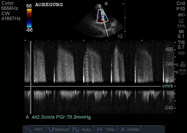
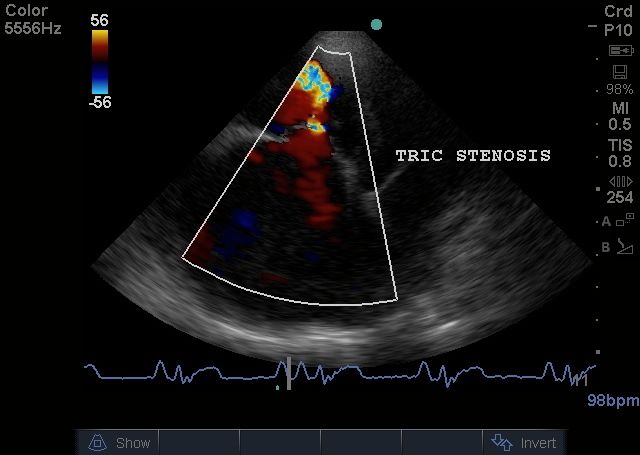
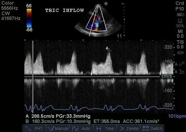
Right Atrial Enlargement in a 9mo Labrador with Tricuspid Dysplasia and Stenosis | Tricuspid stenotic jet into Right Ventricle, same dog | CW Doppler of tricuspid stenosis, same dog |
| Post stenotic dilation of the Aorta in SubAortic Stenosis, 7mo Dogue de Bordeaux | Subcostal View showing peak Aortic velocity of 4m/s | Aortic Insufficiency, same dog, velocity 3m/s. |
| Supracristal VSD in a 6yo Springer Spaniel, right parasternal short axis view | Left to Right PDA in a Shih Tzu cross puppy – CW Doppler of main Pulmonary Artery | Left to right PDA in a Chihuahua puppy |
Right to left PDA in a 9mo Border Terrier – RV hypertrophy and RA enlargement | CW Doppler of markedly elevated tricuspid regurgitation confirming significant Pulmonary Hypertension, same dog | Bubbles in the right heart, bubble study, same dog |
Bubbles in aorta seconds later confirming right to left shunt | Aortic Root Mass in an Irish Wolfhound | |
![13.35.00 hrs __[0001272].jpg](https://lh6.ggpht.com/-xzr7j6C-w6g/Te0GjQ1HY-I/AAAAAAAAAe8/lfUREVlFG54/13.35.00%252520hrs%252520__%25255B0001272%25255D.jpg?imgmax=640)
![13.35.00 hrs __[0001279].jpg](https://lh3.ggpht.com/-xR0q8xgbk40/Te0GjCmp-FI/AAAAAAAAAe4/ZZxJ9M4wpGc/13.35.00%252520hrs%252520__%25255B0001279%25255D.jpg?imgmax=640)
![13.35.00 hrs __[0001270].jpg](https://lh6.ggpht.com/-6UDgRaDRdno/Te0GjMBY78I/AAAAAAAAAe0/2lprSreZI_4/13.35.00%252520hrs%252520__%25255B0001270%25255D.jpg?imgmax=640)
![11.33.32 hrs __[0001375].jpg](https://lh3.ggpht.com/-L-s9Xp_xm00/Te0GidHYCjI/AAAAAAAAAeo/vL5H9iNdrE8/11.33.32%252520hrs%252520__%25255B0001375%25255D.jpg?imgmax=640)