Services
- Abdominal Ultrasound

Patients are positioned in dorsal recumbency (padded trough provided) and all abdominal organs, lymph nodes and vasculature are routinely thoroughly evaluated.
Ectopic ureter and portosystemic shunt assessments are performed only where indicated or specifically requested (see below).
Ectopic Ureter Assessment
Ultrasound assessment of ectopic ureter (ie locating both ureteral insertions on B-mode with confirmation of patency on colour flow) is successful in the vast majority of patients. A 24 – 36 hour fast is required (depending on the age of the patient) to ensure an empty colon, as a full colon will compress the bladder neck.
Thoracic Ultrasound

Ultrasound-guided sampling of mediastinal and thoracic masses is often indicated, for example trucut biopsy of mediastinal or solid lung masses.
In addition guided FNA of diffuse pulmonary interstitial disease has a high diagnostic rate with minimal complications in cases where BAL has been unhelpful, eg diffuse pulmonary interstitial neoplasia, mycoplasma infections.

Ultrasound of the Neck
This is useful for identification of thyroid and parathyroid lesions (hypothyroidism, hypercalcaemic patients).
Echolaryngography is a technique which assesses motion of the arytenoid cartilidges and can be useful for detecting laryngeal paralysis in conscious dogs.
Portosystemic Shunt Evaluation

A full description of vessel origin, course and insertion is provided with associated diameters sufficient for surgical intervention without the requirement for further imaging in the majority of cases.
Ultrasound evaluation of portosystemic shunts holds advantages over CT evaluations such as not requiring anaesthesia or administration of contrast agents with their associated risks, and is also less costly than CT.
Additionally, where a shunt is not located, hepatic trucut biopsy can then be performed to assess for other potential causes of the hepatopathy.
Ultrasound Guided FNA and Biopsy / Coagulation profiling

Ultrasound guided FNA and biopsy are minimally invasive procedures with a high diagnostic rate. Sedation is usually sufficient for FNA and trucut biopsy, depending on the tissue being sampled.
Coagulation profiling (PT and APTT) on-site is available to give immediate results and permit same-day biopsy sampling in patients with questionable coagulability.
PT and APTT values are recommended in all patients undergoing trucut liver biopsy, and in other cases as advised. All patients undergoing trucut biopsy should be hospitalised for the remainder of the day (minimum 3 hours) to monitor for haemorrhage, although this is a very rare complication (less than 0.25%). External laboratory fees are included in the sampling fee.
Echocardiography
Echocardiography includes B-mode, M-mode, and Doppler evaluation (colour flow and spectral pulsed-wave and continuous-wave) of inflow and outflow tracts, using right and left parasternal and subcostal windows. An integrated ECG ensures accurate timing of measurements.
The measurements obtained provide information on cardiac chamber dimensions, wall thicknesses, systolic and diastolic function, filling pressures, and morphology and function of cardiac valves, and allows for the diagnosis of acquired and congenital cardiac diseases.
Breed associated heart testing is not available using NVi services – please go to www.bsava.org.uk/vcs/testing/menu.htm for information on veterinary cardiologists recommended for breed testing schemes.
Endoscopy
Upper and lower gastrointestinal endoscopy are often indicated subsequent to abdominal ultrasound. Please note abdominal ultrasound is considered essential prior to this procedure, and GI endoscopy will never be performed without it (to ensure the entire GI tract is assessed). Pyloric intubation is feasible in the vast majority of patients (including cats) from 3-60kg with our 7.9mm outer diameter, 1.5 metre length, flexible veterinary videoscope. Our large 2.8mm biopsy channel ensures superior biopsy samples, as compared to those obtained via the typical 2mm channel of fiberscopes, which are usually found in general practice.
Bronchoscopy – during this procedure guided BAL is performed using mucus collection traps and suction; this provides a much higher yield than BAL performed using catheter aspiration techniques. Rhinoscopy can be useful as a mobile procedure for retrieval of foreign bodies. However for evaluation of the majority of chronic nasal conditions, referral for CT is advised, where CT will be performed prior to rhinoscopy; this vastly increases the sensitivity of the procedure.
Cystoscopy is a useful procedure for assessment of ureteral insertions, and also to obtain biopsy specimens from bladder or urethral inflammation or neoplasia.


Training
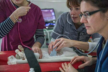
In-house training days can be arranged for ultrasound, ideally on a one to one or one to two basis. Approximately four patients will be required for a day’s training. Using the practices own ultrasound machine will allow for more useable and relevant practical ultrasound training as compared to that obtained by attending an external CPD course, where the equipment used will have capabilities varying significantly from in-house equipment. Additionally, practice revenue generated by performing in-house ultrasound procedures will significantly contribute to covering the cost of the training. In-house endoscopy training is also available.
Training days are also available on Mondays at Westways Vets in Newcastle on a shared two vet per day basis. 4 clinical patients and equipment are provided. This gives an opportunity to attend a local external venue for hands-on ultrasound training without the necessity to arrange your own patients and staff for in-house training, which can be challenging in a busy general practice environment. Patients can be tailored according to training requirements (abdominal vs cardiac).